
CASE STUDYMonitoring brain activity: a wearable brain scanner
WhatIS A WEARABLE BRAIN SCANNER?
Every thought, sensation and movement begins as electrical activity in the brain. As these signals pass between brain cells, they also create tiny magnetic fields. These magnetic fields hold important information about what is happening in the brain, but they are incredibly weak. In fact, they are billions of times smaller than the Earth's magnetic field, which makes them extremely difficult to measure.
Despite their tiny size, these magnetic signals can be measured using a technique called magnetoencephalography (also known as MEG). MEG provides a detailed picture of brain activity with millisecond precision, allowing researchers and clinicians to see when and where activity occurs.
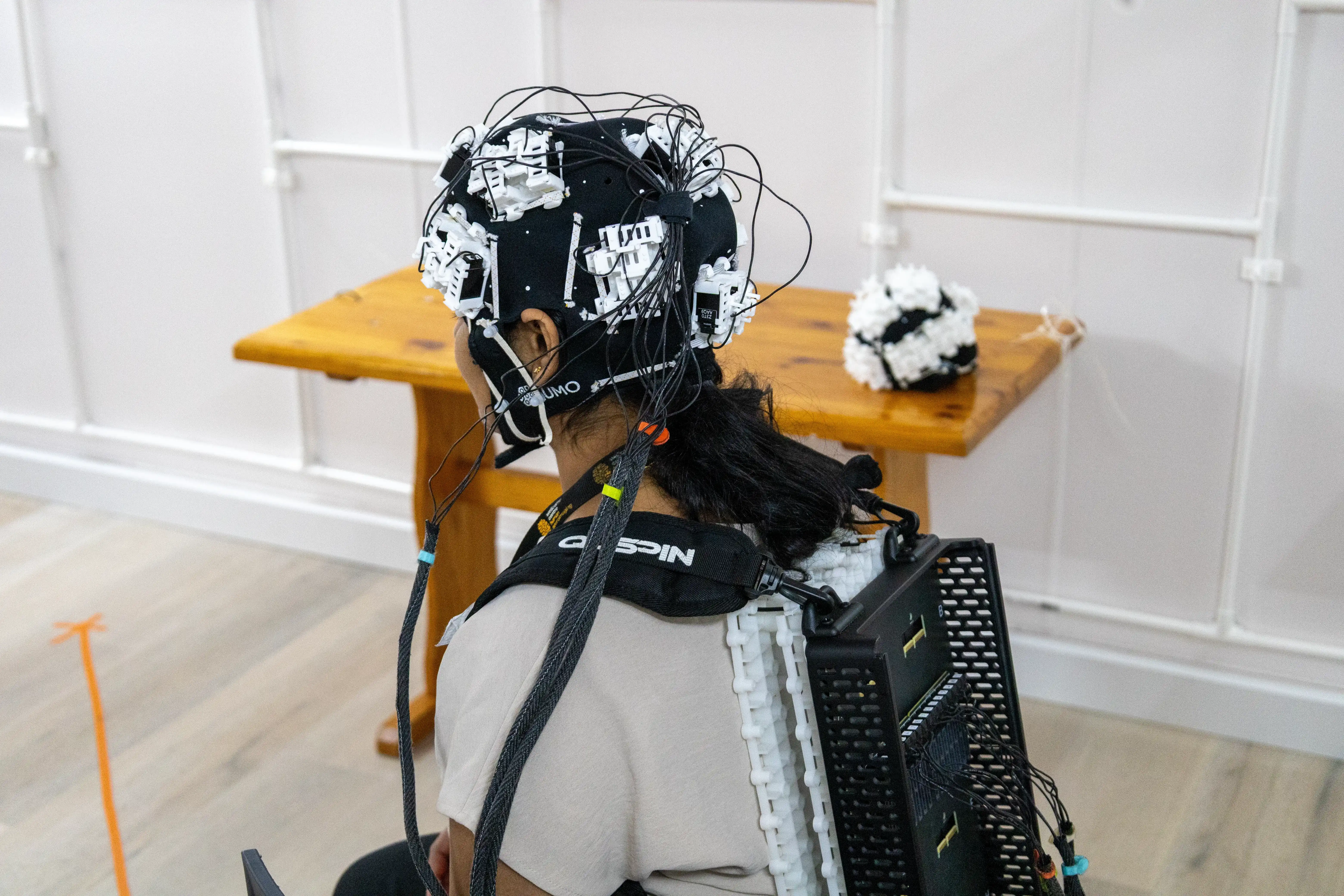
Now, a new generation of quantum sensors called optically pumped magnetometers (OPMs) is transforming this technology. These sensors can be worn directly on the head, creating a wearable form of MEG known as OP-MEG. By allowing people to move naturally during scans, OP-MEG could give us the tools to better understand the brain and improve diagnosis and treatment for a range of neurological conditions.
Traditional MEG systems use highly sensitive sensors called superconducting quantum interference devices (SQUIDs). But these sensors need to be cooled to extremely low temperatures using liquid helium, making the scanners large, expensive and rigid. In fact, because of their size and complexity there are only around 10 of these machines in the UK. In addition, because the sensors sit inside a fixed helmet, patients must remain very still during a scan.
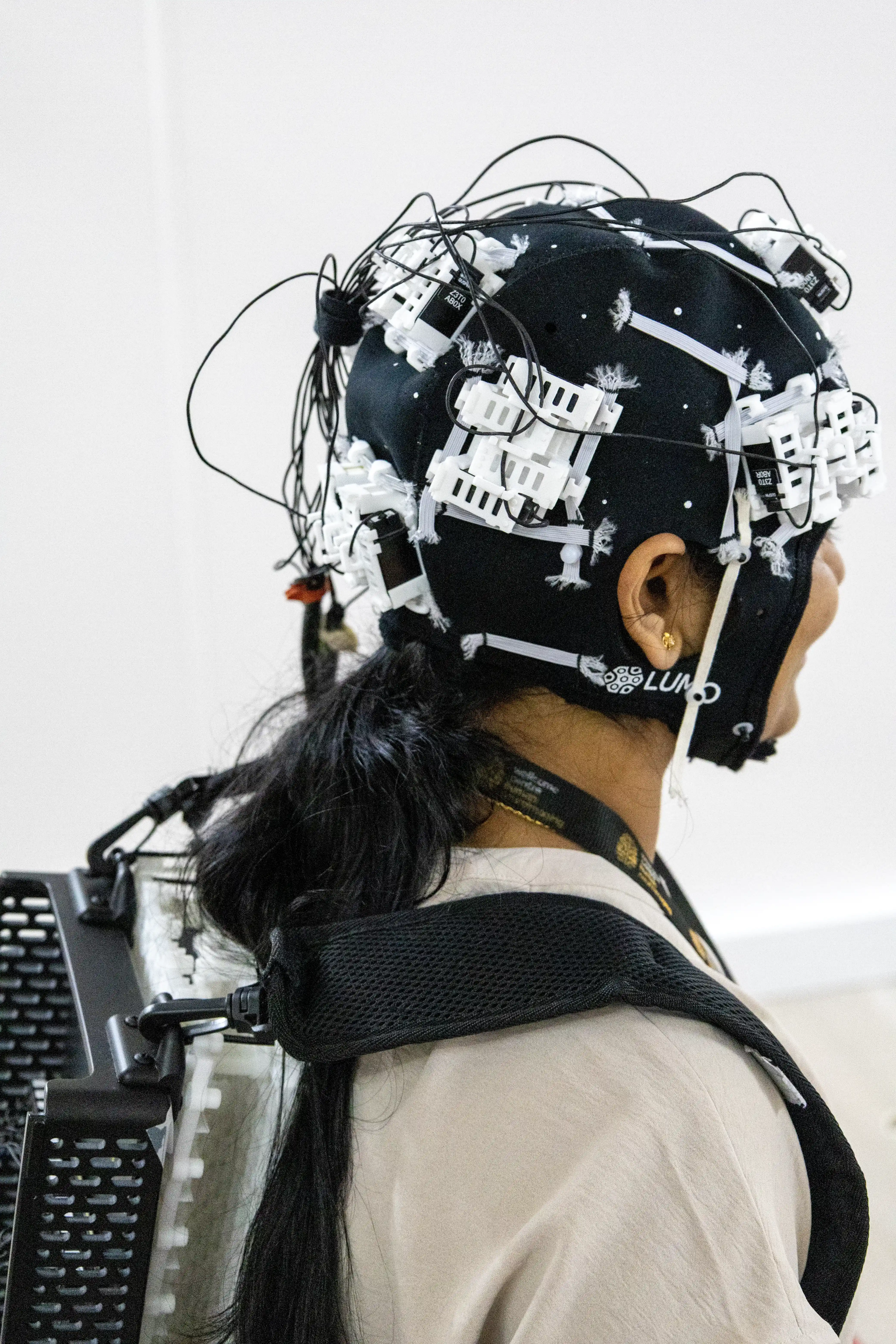
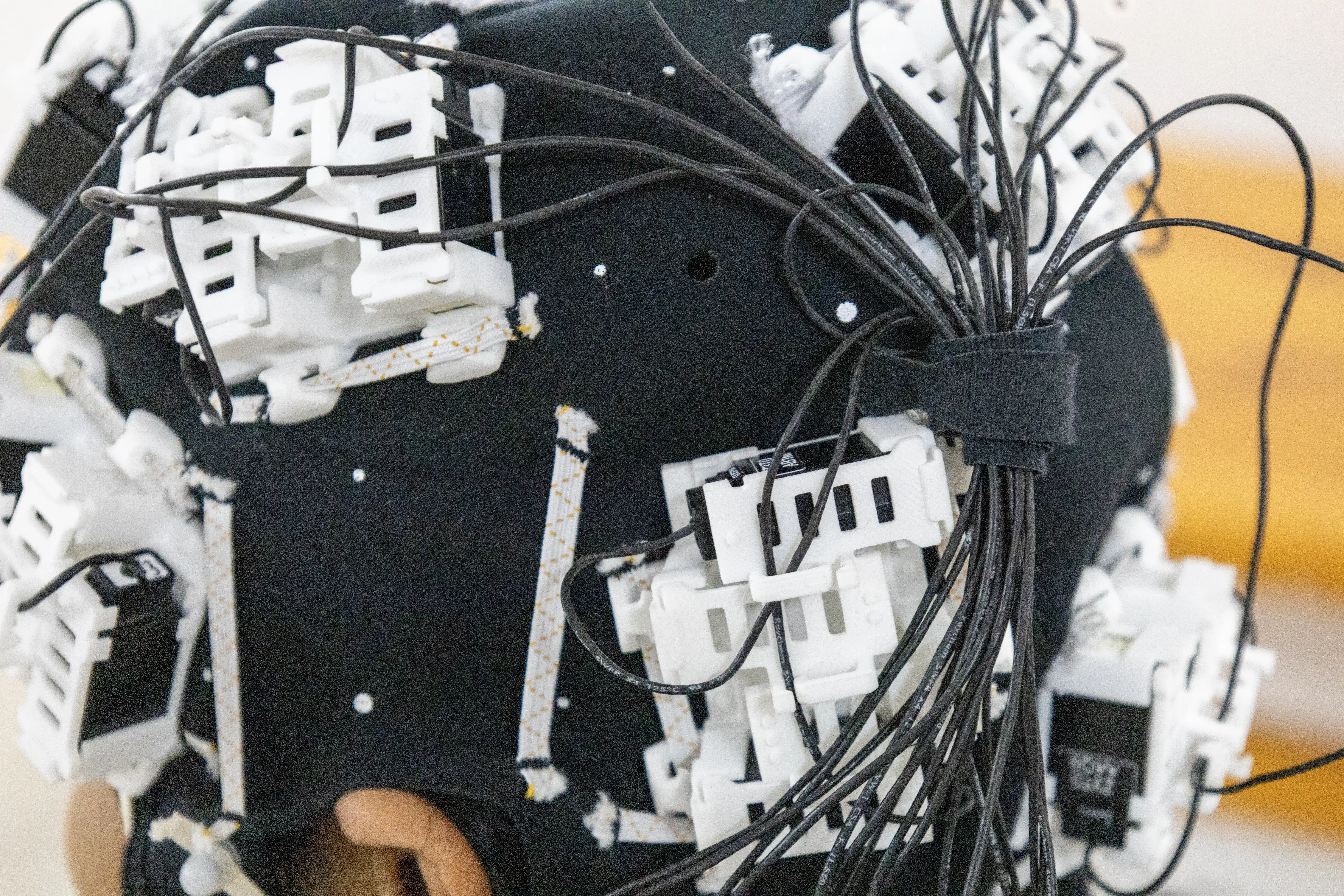
OP-MEG uses much smaller sensors called optically pumped magnetometers. These sensors are roughly the size of a Lego brick and operate at room temperature, which means they can be placed directly on the scalp inside a lightweight wearable helmet.
Because the sensors sit closer to the brain, they can capture stronger signals than traditional MEG systems. Just as importantly, people can move naturally while measurements are being taken. This makes brain imaging more comfortable and practical, particularly for children and people who find it difficult to remain still. It also allows researchers to study brain activity during more natural behaviours, such as moving, interacting with others or potentially even during sleep, opening up new possibilities for both research and healthcare.
Measuring brain activity is useful for a huge variety of conditions, but one of the most promising clinical applications of OP-MEG is in epilepsy.
Epilepsy affects millions of people worldwide and causes repeated seizures. For some patients, medicines do not fully control seizures and surgery may be considered. But for surgery to be successful, clinicians need to identify exactly where in the brain the seizures begin.
This is where OP-MEG comes in. By measuring the magnetic signals produced by abnormal brain activity, it can help clinicians identify the regions where seizures originate. This information can support decisions about whether surgery is appropriate and help surgeons plan procedures more precisely. Researchers hope this could lead to more precise surgery, and ultimately, better outcomes for patients.
The technology may be particularly valuable for children with epilepsy because the wearable sensors allow movement during scanning, making the experience less stressful and enabling higher-quality measurements.
OP-MEG is currently undergoing clinical trials and hospital validation studies, and further evaluation will be needed before it can be adopted routinely in clinical practice.
Many neurological conditions are difficult to diagnose and monitor, creating challenges for both patients and healthcare professionals. To diagnose and treat these conditions effectively, clinicians need better ways to understand how the brain is functioning. OP-MEG combines the precision of advanced brain imaging with the comfort and flexibility of a wearable device. This could make high-quality brain imaging accessible to more people, particularly children and patients who struggle with conventional scans.
By helping us understand the brain in greater detail than ever before, OP-MEG could enable earlier diagnosis, more personalised treatments and entirely new ways of monitoring brain health.
Beyond epilepsy, a particularly exciting application for OP-MEG is in neurodegenerative conditions such as Parkinson's disease and Alzheimer's disease. These conditions affect millions of people worldwide and are becoming increasingly common as populations age, and researchers hope that using OP-MEG to measure subtle changes in brain activity could provide valuable clues for diagnosis, monitoring and treatment.
Beyond brain imaging, researchers are looking at using OPMS for other applications too – like imaging the heart to detect the earliest signs of heart disease.
Because the magnetic signals being measured in the body are so small, one of the biggest drawbacks of MEG is that it needs to be done in a special, magnetically shielded room to prevent other magnetic signals from interfering with the sensors. But researchers are looking into ways to develop new, portable shielding which would help to open up OP-MEG use to a variety of environments.
RESEARCH
Researchers worldwide are driving progress in this area with UK universities and industry playing an important role through the EPSRC-funded Q-BIOMED and QUSIT Hubs.


QBIOMED’s wearable scanner
Close up of QBIOMED’s wearable scanner